New Zealand Doctors Speaking Out with Science
nzdsos.com

The fifth article in our eight-part series on vaccine injuries.

Information

Opinion

Warning

Some may find this content disturbing. The video shows graphic detail from an autopsy.

Vaccine Injuries: Pathology Images

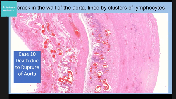
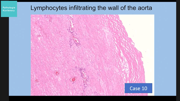
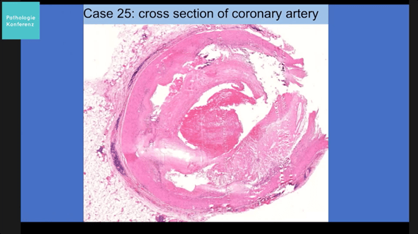
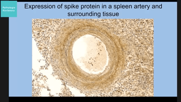
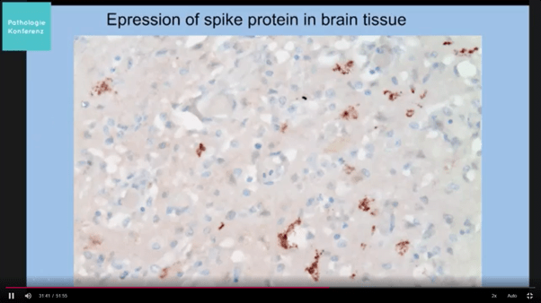
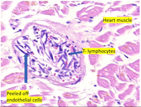
The following are a few images (some potentially disturbing) of what pathologists are seeing worldwide.  It is not at all clear what NZ pathologists are looking for when they examine a person who has died following Covid-19 vaccination.  It is not clear how many post-mortems have been done.  We do not know if they are staining tissues for the presence of spike protein, or whether they are looking for vascular inflammation or micro clotting.

It is also not clear whether biopsies from those still living with various unusual symptoms are being assessed for the presence of spike protein, lipid nanoparticles or synthetic mRNA.

The World Council for Health held a conference to discuss pathology caused by the vaccine.  It can be viewed here and has a number of speakers from various medical, scientific and legal fields presenting.  It was the 6 Feb 2022 session titled ‘Understanding Vaccine Causation Conference’.

This link gives a good explanation of what is happening in the blood vessels post vaccination.

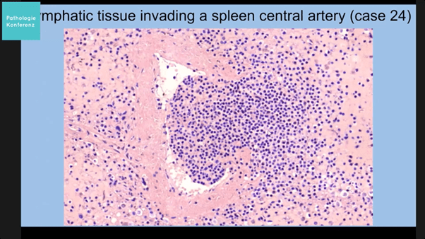
The images below are from Prof Arne Burkhardt Pathology Conference 11 Mar 2022 Germany unless otherwise referenced.

This slide shows inflammation in the main big blood vessel (aorta) in the torso.  The small vessels supplying the muscular artery wall are inflamed (purple areas are numerous inflammatory cells) and the wall itself is inflamed and has ruptured.  Untreated this usually leads rapidly to death from blood loss.
Close up of lymphocyte infiltrates (inflammatory cells) in wall of aorta.  Inflammation may weaken the wall and make it prone to rupture.
Acute thrombosis (blood clot) but also unusual perivasculitis in coronary artery.  This is one of the arteries supplying blood to the heart muscle.  There is inflammation in the wall of this artery (mass of purple cells) and a blood clot has blocked the channel of the artery likely leading to a heart attack.
Spike protein which has been stained brown in spleen artery.  This slide has used a different staining technique and the spike protein itself can be visualised throughout the spleen and one of its arteries. @23 mins
Rupture of central artery in spleen. Lymphocytes (purple dots) which are a type of inflammatory cell are seen invading a burst splenic artery.
Spike protein (stained brown) expressed in heart muscle cells
Spike protein (stained brown) in brain tissue
https://doctors4covidethics.org/vaccine-induced-damage-to-our-vascular-system/ @5.38 epithelial cells sloughed off into the lumen of the blood vessel and inflammatory cells inside the blood vessel

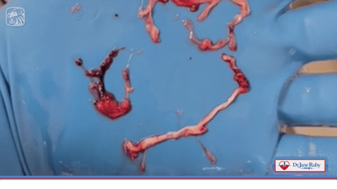
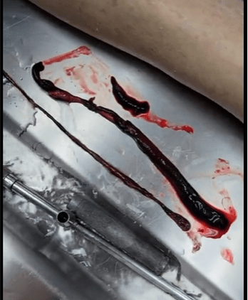
In addition to pathologists, embalmers around the world have been reporting odd things, particularly unusual fibrous blood clots that are obstructing vessels and making it impossible to embalm clients.  These have occurred in both arteries and veins and are different to the clots that sometimes form after death.

Below are long strands of fibrous tissue extracted from vessels during the embalming process, often with blood clots attached to one end suggestive of vaccine injuries.

Unusual long fibrous clots removed from a vein (shorter wider one) and from an artery (longer thinner one) by an embalmer. https://rumble.com/v11qcta-unbelievable-blood-clots-video-from-embalmer-richard-hirschman.html 53 second video removing clots from iliac artery

The New Zealand authorities may be ignoring vaccine injuries but we are not. A subsequent post in this series will focus on what can be done to reduce the effects of vaccine injuries.

Guest Post content does not necessarily reflect the views of the site or its editor. Guest Post content is offered for discussion and for alternative points of view.